Proactive Safety Monitoring in ADC Oncology Trials: Implementing Structured HRCT Surveillance for ILD

Antibody-Drug Conjugates (ADCs) represent one of the most promising frontiers in modern oncology. However, their unique mechanisms of action require equally specialized safety monitoring. Among these considerations, ADC-related Interstitial Lung Disease (ILD) or ADC-induced pneumonitis has emerged as a critical adverse event of special interest.
For clinical trial sponsors, managing ILD risk requires moving beyond standard imaging protocols. To protect patient safety and satisfy regulatory bodies, clinical programs must establish an intentional, expert-led imaging paradigm.
Below, we explore the industry standard for ILD surveillance in ADC trials, the operational requirements for clinical teams, and how specialized central review mitigates study risk.
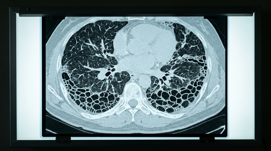
The Gold Standard: Why HRCT is Essential for ADC Safety
In ADC oncology trials, the absolute standard for identifying ILD safety signals is baseline and serial High-Resolution Computed Tomography (HRCT) of the chest. Standard chest CT scans are simply not sufficient; HRCT is the preferred and definitive imaging method for detecting, characterizing, and monitoring the subtle early markers of ILD.
A well-designed ADC clinical protocol typically mandates a structured, multi-phase imaging schedule:
- Baseline Chest Scans: Obtained for every participant before therapeutic treatment to establish a clear anatomical reference point.
- Serial Surveillance: Repeat HRCT scans performed during treatment, typically every 6 to 12 weeks to enable proactive ILD detection.
- Multidisciplinary Integration: Image findings are evaluated alongside clinical symptoms, laboratory results, and pulmonary function tests (PFTs), as imaging findings alone are insufficient to confirm ILD.
Early HCRT Findings in Drug-Induced ILD
When evaluating serial HRCT scans, specialized thoracic radiologists look for distinct patterns that point toward drug-induced pulmonary toxicity. Because treatment‑related ILD can present in diverse and subtle ways, proactive imaging surveillance is designed to identify early abnormalities such as:
- New Ground-Glass Opacities (GGOs): Hazy areas of increased lung attenuation that do not obscure underlying vessels, often representing the earliest signs of inflammation.
- Inflammatory Infiltrates: Areas of consolidation, interlobular septal thickening or fine reticulation reflecting cellular infiltration of the interstitium and alveolar spaces. These key features help distinguish active inflammation from fibrosis
- Organizing Pneumonia-Like Patterns: Pathy peripheral or peribronchial consolidations, reverse-halo configurations, or migratory opacities consistent with an organizing inflammatory response commonly seen in drug-related lung injury.
- Hypersensitivity Pneumonitis-like Patterns: Centrilobular ground-glass nodules, mosaic attenuation and air trapping suggestive of airway centered inflammatory injury.
- Reticular or Mixed Inflammatory Patterns: Fine reticulation, subtle interstitial thickening or combinations for GGO and consolidation that indicate overlapping inflammatory processes
These imaging patterns often overlap with other pulmonary conditions common in oncology patients, including infection, tumor progression, radiation-induced lung injury, and pre-existing ILD, making expert interpretation essential. Because participants are often asymptomatic in the earliest stages, HRCT imaging is often the first and most sensitive indicator of treatment‑related lung toxicity. This is why proactive, serial HRCT surveillance is a cornerstone of modern ADC trial design. Detecting abnormalities during the asymptomatic phase, enables timely protocol-mandated dose holds, reductions or discontinuation, safeguarding the patients’ safety while preserving the integrity of the clinical trial.
Operational Excellence: De-Risking Trials via Central Review
From an operational perspective, the regulatory expectation for ADC programs is exceptionally high. Relying solely on local site radiologists introduces significant variability that can obscure safety signals and compromise study data quality.
To achieve the level of standardization regulators expect, leading ADC programs incorporate:
- Centralized Expert Radiology Review: Dedicated, blinded independent thoracic radiologists apply uniform criteria across all sites, ensuring consistent interpretation of HRCT findings.
- Joint Adjudication Committees: Structured workflows where expert radiologists and independent clinicians collaboratively review borderline or complex cases to reach a consensus determination.
The Voiant Advantage
As an industry-leading imaging core lab, Voiant delivers the specialized therapeutic expertise, calibrated operational workflows, and low-latency digital infrastructure required for seamless HRCT surveillance. Centralizing imaging with Voiant ensures that subtle ILD safety signals are detected early, interpreted consistently, and aligned with regulatory expectations.
Practical Takeaway for Trial Designers
If you are designing or evaluating an ADC oncology trial, imaging cannot be reduced to “a chest CT”. Success requires a structured HRCT surveillance program that includes rigorous baseline imaging, scheduled follow-up scans, adherence to standardized acquisition guidelines and expert central interpretation. This is the operational foundation that protects patients and preserves the integrity of your trial.
Discover how Voiant helped an ADC trial enhance ILD detection through an integrated imaging workflow
Want more like this? Join our mailing list.
Gain insights from the imaging experts behind 70+ successful drug approvals trusted by leading biopharma teams worldwide.
"*" indicates required fields
References
- Chan KL, Faiz SA, Altan M, et al. Updates in drug-related pneumonitis due to targeted oncologic therapies. J Immunother Precis Oncol. 2024;7(4):272-282. https://doi.org/10.36401/JIPO-24-12
- Distefano G, Fanzone L, Palermo M, et al. HRCT patterns of drug-induced interstitial lung diseases: a review. Diagnostics (Basel). 2020;10(4):244. https://doi.org/10.3390/diagnostics10040244
- Lederer C, Storman M, Tarnoki AD, et al. Imaging in the diagnosis and management of fibrosing interstitial lung diseases. Breathe (Sheff). 2024;20(1):240006. https://doi.org/10.1183/20734735.0006-2024
- Podolanczuk AJ, Hunninghake GM, Wilson KC, et al. Approach to the evaluation and management of interstitial lung abnormalities: an official ATS clinical statement. Am J Respir Crit Care Med. 2025;211(7):1132-1155. https://doi.org/10.1164/rccm.v211erratum4
- Voiant Clinical. Integrated RECIST + ILD workflow enhances safety monitoring in an ADC oncology program. https://www.voiantclinical.com/resources/case-studies/integrated-recist-ild-workflow-enhances-safety-monitoring-in-an-adc-oncology-program/
- Voiant Clinical. Respiratory therapeutic area expertise. https://www.voiantclinical.com/therapeutic-areas/respiratory/